We use cookies to enhance the usability of our website. If you continue, we'll assume that you are happy to receive all cookies. More information. Don't show this again.
DACT2
HPA
RESOURCES
  • TISSUE
  • BRAIN
  • SINGLE CELL
  • SUBCELLULAR
  • CANCER
  • BLOOD
  • CELL LINE
  • STRUCTURE & INTERACTION
ABOUT
  • INTRODUCTION
  • HISTORY
  • ORGANIZATION
  • PUBLICATIONS
  • ANTIBODY SUBMISSION
  • ANTIBODY AVAILABILITY
  • ACKNOWLEDGMENTS
  • CONTACT
NEWS
  • NEWS ARTICLES
  • PRESS ROOM
LEARN
  • DICTIONARY
  • PROTEIN CLASSES
  • PROTEIN EVIDENCE
  • METHODS
  • EDUCATIONAL VIDEOS
DATA
  • DOWNLOADABLE DATA
  • PUBLICATION DATA
  • RELEASE HISTORY
HELP
  • ANTIBODY VALIDATION
  • ASSAYS & ANNOTATION
  • DISCLAIMER
  • HELP & FAQ
  • PRIVACY STATEMENT
  • LICENCE & CITATION
Fields »
Search result

Field
Term
Gene name
Class
Subclass
Class
Keyword
Chromosome
External id
Tissue
Cell type
Expression
Antibody panel
Tissue
Main location
Patient ID
Annotation
Tissue
Category
Tau score
Cluster
Reliability
Brain region
Category
Tau score
Brain region
Category
Tau score
Brain region
Category
Tau score
Cluster
Reliability
Tissue
Cell type
Enrichment
Cell type
Category
Tau score
Cell type
Category
Tau score
Cell type
Category
Tau score
Cell lineage
Category
Tau score
Cluster
Cluster
Location
Searches
Location
Cell line
Class
Type
Phase
Reliability
Cancer
Prognosis
Cancer
Category
Cancer
Category
Tau score
Cluster
Variants
Interacting gene (ensg_id)
Type
Number of interactions
Pathway
Category
Score
Score
Score
Validation
Validation
Validation
Validation
Antibodies
Data type
Column


  • SUMMARY

  • TISSUE

  • BRAIN

  • SINGLE CELL

  • SUBCELL

  • CANCER

  • BLOOD

  • CELL LINE

  • STRUCT & INT

  • DACT2
BRAIN TISSUES Show tissue menu
AMYGDALA BASAL GANGLIA CEREBELLUM CEREBRAL CORTEX CHOROID PLEXUS HIPPOCAMPAL FORMATION HYPOTHALAMUS MEDULLA OBLONGATA
MIDBRAIN OLFACTORY BULB PITUITARY GLAND PONS RETINA SPINAL CORD THALAMUS WHITE MATTER
PRIMARY MOUSE DATA
ANTIBODIES
AND
VALIDATION
Brain proteome
MOUSE BRAIN TISSUE
Gene description Dishevelled binding antagonist of beta catenin 2
Mouse gene ENSMUSG00000048826 (version 10)
Annotation summary DACT2 RNA transcripts are highly distributed in placenta, in moderate levels in brain and lower in the rest of human organs. In mouse brain, available transcriptomic and proteomic data agree on an enrichment across cortical areas, demonstrating a predominantly dendritic/synaptic staining pattern. Indeed, moderate-intensity diffuse/synaptic-type immunoreactivity is noticed throughout the entire forebrain. In the brainstem, this patterns is rather restricted to certain gray matter areas, like the periaqueductal gray matter, dorsal raphe, substantia nigra, dorsal part of medulla around the fourth ventricle. In addition, moderate-strong somato-dendritic immunostaining of neurons is noticed in the lateral septum, nucleus accumbens, cerebellar Purkinje-cells and in multiple brainstem nuclei. In addition, distinct strong axonal immunostaining is noticed in the olfactory bulb glomerular- and external plexiform layers.
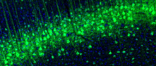
Positive cells and structuresi

Manually selected location of the protein positivity, observed by immunofluorescence staining in mouse brain.

Soma, dendrite, axon and synapse in neurons.
FRONT BACK
Midbrain
Pons
Hippocampal Formation
Protein
Relative fluorescence intensityi

Protein expression in mouse brain using immunofluorescence is reported as mean fluorescence intensity .

Mouse brain region
255
0
Cerebral cortex
Frontal association cortex (fra)
Motor cortex (m)
Cingulate cortex (cg)
Piriform cortex, L1 (pirl1)
Piriform cortex, L2 (pirl2)
Piriform cortex, L3 (pirl3)
Insular cortex (i)
Somatosensory cortex (s)
Retrosplenial granular cortex (rsg)
Parietal association cortex (p)
Entorhinal cortex (ent)
Visual cortex (v)
Olfactory bulb
Anterior olfactory nucleus (aon)
Granule cell layer (gro)
Internal plexiform layer (ipl)
Mitral cell layer (mi)
Glomerular layer (gl)
Rostral migratory stream (rms)
External plexiform layer (epl)
External plexiform layer of the accessory OB (epla)
Granule cell layer of the accessory OB (gra)
Glomerular layer of the accessory OB (gla)
Hippocampus
Polymorph layer of the dentate gyrus (podg)
Molecular layer of the dentate gyrus (modg)
Granular dentate gyrus (grdg)
CA1 - oriens layer (ca1or)
CA1 - pyramidal layer (ca1py)
CA1 - radiatum layer (ca1ra)
CA2 - oriens layer (ca2or)
CA2 - pyramidal layer (ca2py)
CA2 - radiatum layer (ca2ra)
CA3 - oriens layer (ca3or)
CA3 - pyramidal layer (ca3py)
CA3 - radiatum layer (ca3ra)
Stratum lucidum (slu)
Lacunosum moleculare (lmol)
Subiculum (sub)
Amygdala
Nucleus of the lateral olfactory tract (lot)
Basal medial amygdaloid nucleus (bma)
Basal lateral amygdala* (*bla) N/A
Basal lateral amygdaloid nucleus (bla)
Cortical amygdala (aco)
Central amygdala (ce)
Medial amygdaloid nucleus (mea)
Thalamus
Medial geniculate nucleus (mg)
Parafascicular thalamic nucleus (pf)
Pregeniculate nucleus (pg)
Stria terminalis (st)
Zona incerta (zi)
Anterodorsal thalamic nucleus (ad)
Reticular thalamic nucleus (rt)
Vental anterior thalamic nucleus (va)
Medial habenular nucleus (mhb)
Laterodorsal thalamic area (ld)
Paraventricular thalamic nucleus (pv)
Central medial thalamic area (cm)
Ventral lateral thalamic area (vl) N/A
Ventral medial thalamic area (vm)
Lateral habenulal nucleus (lhb)
Ventral posterior thalamus (vpt)
Anterior pretactal nucleus (apt)
Retromammillary nucleus (rm)
Hypothalamus
Dorsal tuberomammillary nucleus (dtm)
Mammillary nucleus (mn)
Periventricular hypothalamic nucleus (pe)
Supraoptic nucleus (so)
Tuberal nucleus (tu)
Ventral tuberomammillary nucleus (vtm)
Lateral preoptic area (lpo)
Medial preoptic area (mpo)
Suprachiasmatic nucleus (sch)
Paraventricular hypothalamic nucleus (pa)
Anterior hypothalamic area, central (ahc)
Ventral medial hypothalamic nucleus (vmh)
Ventral medial hypothalamic nucleus* (*vmh) N/A
Arcuate nucleus (arc)
Arcuate nucleus* (*arc) N/A
Peduncular part of lateral hypothalmus (plh)
Dorsal medial hypothalamic nucleus (dm)
Stria terminalis* (*st) N/A
Cerebellum
Vestibulocerebellar nucleus (vecb) N/A
Moleuclar layer of the cerebellum (cemol)
Purkinje layer of the cerebellum (cepur)
Granular layer of the cerebellum (cegr)
Pons
Barrington's nucleus (bar) N/A
Koelliker-fuse nucleus (kf) N/A
Motor tregiminal nucleus (5n)
Parabrachial nucleus (pbp)
Principle sensory trigeminal nucleus (pr5)
Locus coeruleus (lc)
Pontine nucleus (pn)
Vestibular nucleus (ve)
Pontine reticular nucleus, oral (pno)
Lateral lemniscus (ll)
Superior paraolivary nucleus (spo)
Medulla oblongata
Nucleus of the solitary tract (sol)
Raphe magnus nucleus (rmg)
Cochlear nucleus (cn)
Lateral paragigantocellular nucleus (lpg)
Raphe pallidus nucleus (rpa)
Facial nucleus (7n)
Hypoglossal nucleus (12n)
Ambiguus nucleus (amb)
External cuneate nucleus (ecu)
Inferior olivary nucleus (io)
Raphe obscures nucleus (rob)
Dorsal motor nucleus of vagus (10n) N/A
Circumventricular organs
Subcommissural organ (sco)
Subfornical organ (sfo)
Median eminence (me)
Medulla (ap) N/A

Antibodies in assayi

The internal antibody ID for the antibody/antibodies available for this specific target, click link for more antibody information.

HPA030251

Contact

  • NEWS ARTICLES
  • PRESS ROOM

The Project

  • INTRODUCTION
  • ORGANIZATION
  • PUBLICATIONS

The Human Protein Atlas

  • DOWNLOADABLE DATA
  • LICENCE & CITATION
  • HELP & FAQ
The Human Protein Atlas project is funded
by the Knut & Alice Wallenberg Foundation.


contact@proteinatlas.org